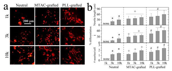
Figure 5.
PC12 cell differentiation on the hydrogels. (a) Fluorescent images of PC12 neurites induced by NGF at day 7. Stained with rhodamine-phalloidin. Scale bar of 200 μm is applicable to all. (b) Neurite length, percentage of differentiated cells, and the number of neurites per cell for PC12 cells at day 7. *: p < 0.05 between two marked samples. +,#: p < 0.05 between two marked samples and relative to neutral hydrogels made from the same PEGDAs.

