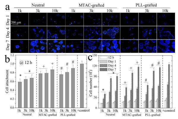
Figure 6.
NPC proliferation on the hydrogels. (a) Nuclei (blue) of NPCs for 1, 4, and 7 days. Scale bar of 200 μm is applicable to all. (b) NPC attachment at 12 h post-seeding. Positive (+) control: TCPS. (c) NPC cell number at 12 h, days 1, 4, and 7 post-seeding. *: p < 0.05 between two marked samples at the same time. +,#: p < 0.05 between two marked samples and relative to the neutral hydrogels made from the same PEGDAs at the same time. Positive (+) control: TCPS.

