
Figure 4.
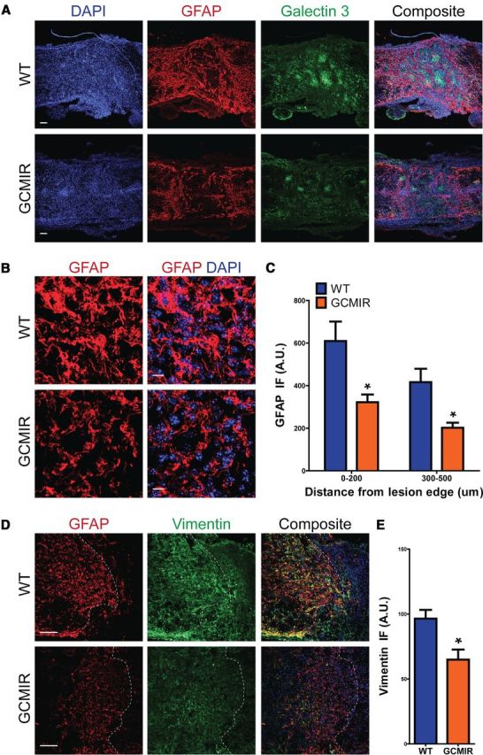
miR-21 attenuates astrocytic hypertrophy 14DPI after SCI. WT and GCMIR21 mice were analyzed 14 DPI after SCI. A, Longitudinal cross-sections revealed that astrocytes were less hypertrophic and expressed less GFAP (red) in GCMIR21 mice compared with WT. Compaction by astrocytes of activated microglia marker Galectin 3+ (green) was also reduced in GCMIR21 mice compared with WT. Scale bar, 100 μm. B, Magnified images of astrocytes adjacent to lesion showed smaller astrocytes with thinner processes in GCMIR21 mice compared with WT. (GFAP, red; DAPI, blue.) Scale bar, 10 μm. C, Quantification of GFAP immunofluorescence (IF; arbitrary units, A.U.) 0–200 μm and 300–500 μm away from lesion edge. D, Vimentin immunofluorescence (green) is also reduced in astrocytes adjacent to injury site in GCMIR21 mice compared with WT. Dashed white lines indicate lesion edge. Scale bar, 100 μm. E, Quantification of vimentin staining within 200 μm of injury site. n = 4, *p < 0.05 by Student's t test.