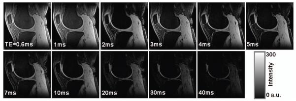
Fig. 3.
Images of a subject across the 11 echo times (TEs), displayed at the same window/level. Signal intensity in the deep layer of cartilages decreased faster with TE increasing than in the superficial layers, due to faster T2* relaxation. SNR on the patellar cartilage is 87, 92, 81, 71, 66, 59, 47, 43, 29, 23, and 16 for TE=0.6-40ms, respectively. (3T, FOV=140mm, matrix=256, thickness=2mm, TR=80ms, θ=30°)

