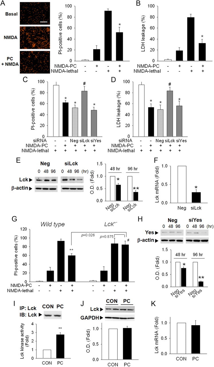
Figure 1.

Lck mediates PC neuroprotection against NMDA-induced cytotoxicity in primary cortical neurons. A, PC with NMDA (10 μm) decreased vulnerability to lethal levels of NMDA (50 μm). Neurons were stained with PI at 24 h after lethal NMDA exposure. Scale bar, 200 μm. B, Lactate dehydrogenase (LDH) released into the medium was measured. C, D, Transfection of siRNA against Lck but not Yes reversed PC neuroprotection in PI-staining (C) or LDH leakage (D) at 24 h after lethal NMDA exposure. Neg, Nontargeting negative siRNA; siYes, siRNA against Yes. *p < 0.05 versus NMDA lethal; #p < 0.05 versus Neg+NMDA PC+NMDA lethal. E, Lck protein levels were significantly decreased after siLck transfection. β-actin, Loading control. F, Lck mRNA was reduced at 48 h after siLck transfection. *p < 0.05, **p < 0.01 versus Neg. G, NMDA PC neuroprotection was not observed in cortical neurons from Lck−/− mice. **p < 0.01 versus NMDA lethal; #p < 0.05 versus wild-type NMDA PC+NMDA lethal. H, siYes transfection decreased Yes protein level. I, Lck kinase activity was significantly increased after NMDA PC. Cell lysates were immunoprecipitated with Lck Ab and applied to kinase assay. Total level of Lck in each immunoprecipitates was determined by Western blot. *p < 0.05 versus control without PC; #p < 0.05 versus NMDA PC. J, NMDA PC did not change total Lck protein levels. GAPDH, A loading control. K, Gene transcription levels of Lck were not affected by NMDA PC. A, n = 6; B, I, n = 4; C–H, K, n = 3; J, n = 4–5. All values are means ± SEM and analyzed by Student's t test.
