Figure 2.
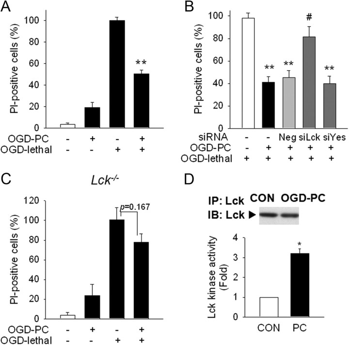
Lck is important in OGD PC neuroprotection. A, Primary cortical neurons were incubated in glucose free Earl's balanced salt solution under 5% CO2/95% N2 at 37°C for OGD. OGD PC (30 min) reduced lethal OGD (2 h)-induced neuronal cytotoxicity in PI-staining. OGD PC was performed 24 h before lethal OGD stimulation. Cytotoxicity was examined at 24 h after lethal OGD stimulation. B, Silencing Lck significantly reversed OGD PC neuroprotection. siRNAs were transfected 48 h before OGD PC, and OGD PC/lethal OGD exposure was performed as described above. *p < 0.05, **p < 0.01 versus OGD lethal; #p < 0.05 versus Neg+OGD PC+OGD lethal. C, Protective effect of OGD PC was not found in primary neurons from Lck−/− mice. D, Lck kinase activity was increased during OGD PC. Neuronal lysates were collected after OGD PC and used for Lck immunoprecipitation and kinase assay. Total level of Lck in each immunoprecipitates was determined by Western blot. *p < 0.05 versus control without PC. A, n = 4; B, D, n = 3; C, n = 5. All values are means ± SEM and analyzed by Student's t test.

