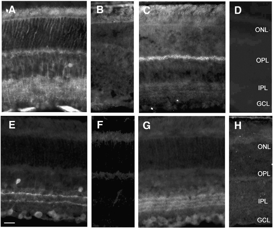
Fig. 1.
VGCC β subunit distribution in wild type and β subunit null mouse retina. A) β1 is present on cell membranes in the middle portions of the INL and processes extending into the ONL and IPL; lateral processes can be detected; B) no β1 is found in CNS β1 null retinas; C) β2 is expressed as punctate label in the OPL with weak but punctate label throughout the IPL; D) no β2 is present in CNS β2 null retinas; E) β3 is seen in two distinct narrow bands in the IPL, scattered cells throughout the most proximal row of the INL, and some cells in the GCL; The strongest immunoreactivity is observed in the outermost band of the IPL; F) no β3 is noted in β3 null retinas; G) β4 is distributed in 4 bands in the IPL H) no β4 is detected in β4 null retinas. Scale bar = 25 µm.

