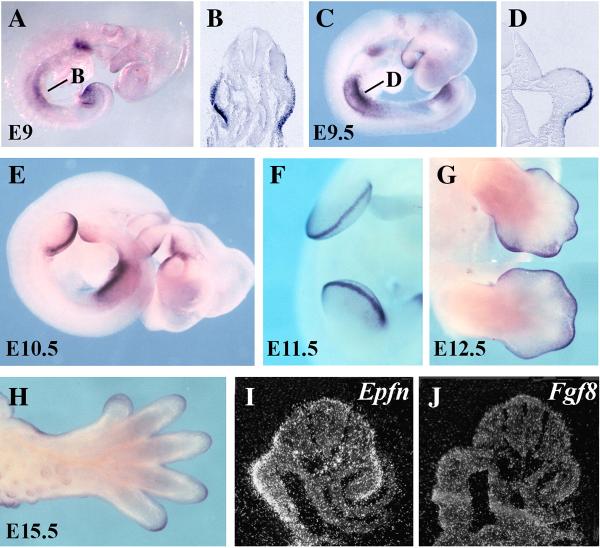
Figure 1. Expression pattern of Epiprofin during mouse limb bud development.
Whole-mount in situ hybridization was performed on wild-type embryos of the indicated age with an Epfn antisense riboprobe. (A–D) At E9 and E9.5 Epfn is expressed in the presumptive fore- and hindlimb ectoderm as shown in the whole mounts and corresponding sections at the forelimb level as marked in the corresponding whole mount (B, D). At E10.5 (E) and E11.5 (F) Epfn expression is predominantly confined to the AER. At E12.5 (G) Epfn expression continues in the AER and extends into the dorsal and ventral ectoderm particularly at digital tips. At 14.5 (H) Epfn expression still occurs in the ectoderm of the digital tips. (I, J) show two consecutive serial sections (6 mm apart) through the presumptive forelimb of an E9 mouse embryo, showing Epfn expression in the limb field ectoderm (I), while Fgf8 expression has not yet been activated (J) at this stage.

