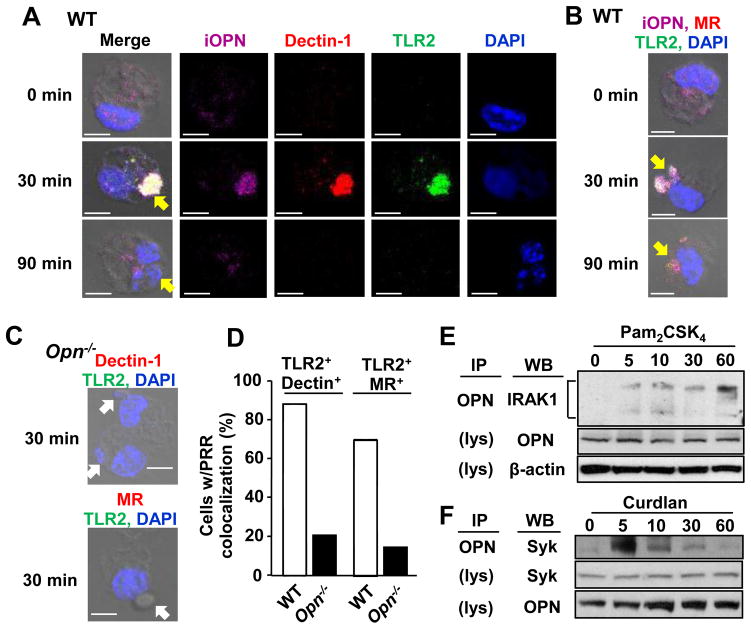
Figure 2. Interaction of iOPN and fungal PRRs by Pneumocystis treatment.
(A) Representative confocal microscopic images of OPN (magenta), dectin-1 (red), TLR2 (green) and DAPI (blue) staining in WT macrophages at indicated time points after co-culture with Pneumocystis. (B) Same as (A), except that MR is indicated with red. (C) No distinct cluster formation of PRRs in Opn−/− macrophages 30 min after Pneumocystis co-culture. (A–C) Yellow and white arrows represent phagocytosed Pneumocystis and Pneumocystis contacting to macrophages, respectively. Scale bars = 5 μm. See Supplemental Fig. 2A and 2B for single color staining images for (B) and (C), respectively. (D) Calculated frequency of macrophages with PRR colocalization (TLR2 and dectin-1 or MR) at 30 min after Pneumocystis co-culture. (E) Immunoprecipitation with OPN in RAW 264.7 cells stimulated with Pam2CSK4 (100 ng/ml) for 0, 5, 10, 30, and 60 min. Immunoblot were performed with anti-IRAK-1. OPN and β-actin were detected in non-immunoprecipitated cell lysates (indicated as “lys”). (F) Co-immunoprecipitation of iOPN and Syk in peritoneal macrophages stimulated with curdlan (100 μg/ml).

