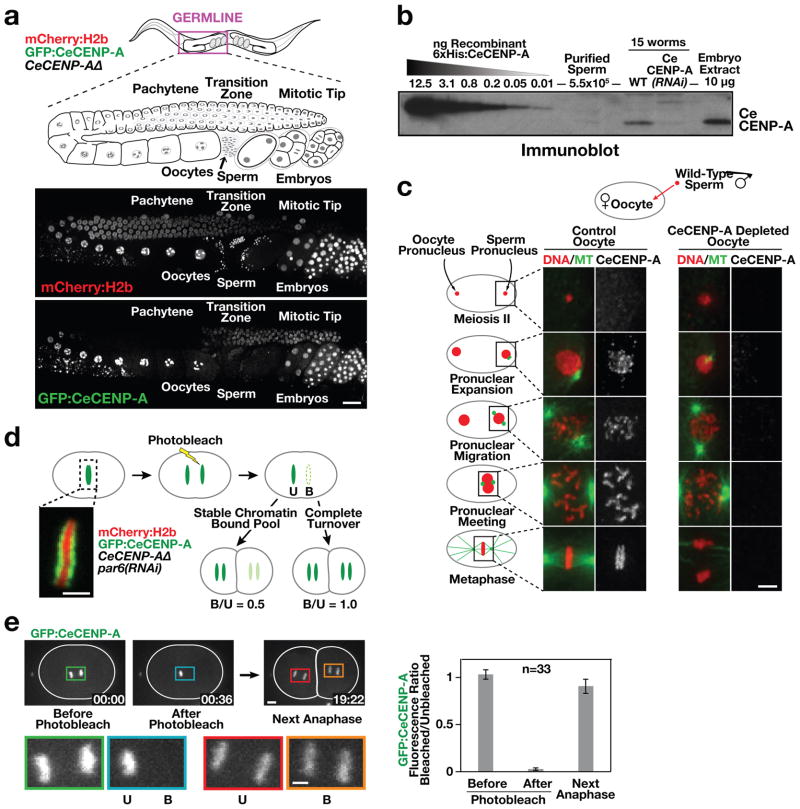
Figure 1. CeCENP-A dynamics in meiotic prophase, at fertilization and across embryonic divisions.
a, Gonad region of an adult hermaphrodite co-expressing GFP–CeCENP-A and mCherry–histone H2b in a CeCENP-AΔ (hcp-3(ok1892; see also Supplementary Figs 1 and 2) background (see also Supplementary Figs 1 and 2). Scale bar, 20 μm. b, Quantitative immunoblot showing that sperm lack a significant pool of CeCENP-A (see also Supplementary Fig. 2c, d). c, Fertilized one-cell control or CeCENP-A-depleted embryos at different stages of the first mitotic division were immunostained for CeCENP-A and α-tubulin (MT). Wild-type (N2) males were mated to fem-1 mutant worms to ensure all embryos were cross-progeny. Scale bar, 5 μm. d, Schematic of photobleaching experiment to assay CeCENP-A inheritance across early embryonic divisions. par-6 RNA interference (RNAi) abolishes developmental asynchrony in the two-cell embryo. Unbleached (U) and bleached (B) chromatid sets are indicated. Scale bar, 2 μm. e, Representative images and quantification of the photobleaching experiment. Higher magnification views highlight bleached and unbleached chromatid sets. Error bars are 95% confidence intervals for the means. Scale bars, 5 μm.

