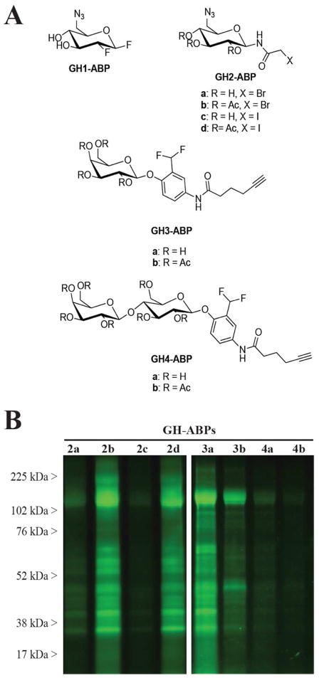
Figure 1.
(A) Structures of glycoside hydrolase-directed activity- based probes (GH-ABPs). (B) C. thermocellum secretome samples, containing cellulosome proteins, were incubated at 1 mg/mL with individual GH-ABPs (75 μM). Click chemistry was used to append the fluorophores rhodamine-azide and - alkyne, and proteins were separated by SDS-PAGE and imaged to reveal the fluorescent ABP-labeled proteins. Control gels and protein abundance stains are shown in the Supporting Information.

