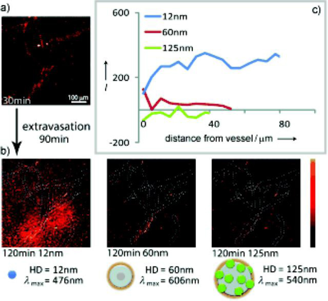
Fig. 3.
Real-time intravital imaging of size-dependent QDs distribution in a SCID mouse bearing a Mu89 melanoma. (a) Multiphoton microscopy image of the distribution of NPs at 30 min. (b) Multiphoton microscopy images of distribution of the NPs in the same region as (a) at 120 min post-injection. (c) Penetration depth analysis at 60 min post-injection. (Reproduced with permission from [14], copyright by the John Wiley and Sons)

