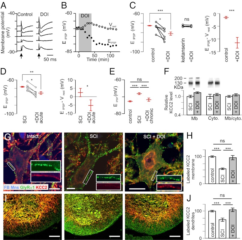
Fig. 1.
Activation of 5-HT2Rs hyperpolarizes EIPSP and increases membrane expression of KCC2. (A) IPSPs evoked by stimulation of the ventral funiculus of the spinal cord (arrow) at different holding potentials in a motoneuron from a P6 intact rat before and after adding DOI (10 µM). (B) Time course of the change in EIPSP and Vrest. (C) EIPSP and driving force (EIPSP-Vrest) measured in control conditions (n = 21) and after adding DOI (n = 8). ***P < 0.001 (Mann–Whitney test). Six motoneurons were tested before and after DOI. *P < 0.05 (Wilcoxon paired test). The effects of DOI were prevented in the presence of ketanserin (10 µM; n = 4) (Center). (D) Effects of DOI (1–1.5 µM) on EIPSP and driving force in motoneurons recorded 4–6 d after neonatal SCI (17 and 6 cells recorded in the absence and the presence of DOI, respectively). **P < 0.01, *P < 0.05 (Mann–Whitney test); *P < 0.05 (Wilcoxon paired test). (E) EIPSP was significantly more hyperpolarized after chronic treatment of DOI (n = 9) compared with SCI animals (n = 22) but not different compared with control animals (n = 21). ns, not significant (P > 0.05); ***P < 0.001 (one-way ANOVA, Tukey’s post tests). (F, Upper) Western blots of membrane and cytoplasmic fractions of lumbar spinal cords labeled with a KCC2-specific antibody. The 130- to 140-kDa and the >200-kDa bands correspond to the monomeric and oligomeric proteins, respectively (53). (F, Lower) Quantification of KCC2 expression in DOI-treated rats after neonatal SCI (percentage of untreated rats). *P < 0.05 (Mann–Whitney test; n = 6 in each group). (G) Dual labeling of GlyRα1 and KCC2 on FB-labeled ankle extensor motoneurons, in three conditions (P7): intact, neonatal SCI, and chronic DOI treatment. (Scale bars: 10 µm.) (H) Quantification of the density of membrane KCC2 labeling (ratios of labeled pixel surface per somatic perimeter) in intact rats (n = 34 motoneurons) and untreated (n = 41) or DOI-treated (n = 44) rats with neonatal SCI (three rats in each group). ***P < 0.001 (Kruskal–Wallis test, Dunn’s post tests). (I) Ventral part of the lumbar spinal cord exhibiting KCC2-immunopositive dendrites stretching out through the white matter. (Scale bars: 100 µm.) (J) Quantification of the KCC2 labeling in the white matter in intact rats (n = 34) and untreated (n = 41) or DOI-treated (n = 44) rats with neonatal SCI. ***P < 0.001 (Kruskal–Wallis test, Dunn’s post tests).

