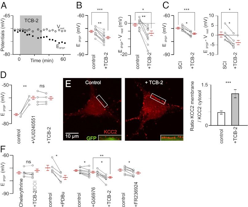
Fig. 2.
Involvement of 5-HT2ARs via a PKC-dependent signaling pathway. (A) TCB-2 (0.1 µM; 30 min) induces a hyperpolarizing shift of EIPSP without concomitant effect on Vrest (P6). (B, Left) Effect of TCB-2 (0.1 µM, gray; 10 µM, black) on 10 motoneurons recorded from control rats (P5–P6). ***P < 0.001 (Mann–Whitney test; n = 12 and n = 14 before and under TCB-2, respectively); **P < 0.01 (Wilcoxon paired test). The difference was also significant when considering only the effect of the lowest concentration [0.1 µM; n = 6; *P < 0.05 (Wilcoxon paired test)]. (B, Right) Effect of TCB-2 (0.1–10 µM) on the driving force (control, n = 12; TCB-2, n = 14). *P < 0.05 (Mann–Whitney test); **P < 0.01 (Wilcoxon paired test). (C) Effect of TCB-2 (0.1 µM) on EIPSP and the driving force of motoneurons (six and nine in the absence and in the presence of TCB-2, respectively) in animals with SCI at birth. ***P < 0.001, *P < 0.05 (Mann–Whitney test); *P < 0.05 (Wilcoxon paired test) (n = 6). (D) EIPSP was significantly depolarized by the application of the KCC2 blocker VU0240551 (25 µM), which prevented the hyperpolarizing effect of TCB-2 (0.1 µM; n = 11). Averages are taken from 3 motoneurons in control situation, 9 motoneurons under VU0240551, and 11 motoneurons in the presence of both VU0240551 and TCB-2. ns, P > 0.05; **P < 0.01 (Mann–Whitney test). (E) TCB-2 (0.1 µM; 30 min) significantly increased the expression of KCC2 at the plasma membrane of Hb9::eGFP transgenic motoneurons in culture (14 DIV). (E, Left and Center) Merge images (Insets) show the KCC2 immunolabeling at the periphery of GFP-expressing cytosol. (E, Right) Ratio of KCC2 expression between plasma membrane and cytosol. ***P < 0.001 (t test; n = 34 cells from two experiments analyzed in each condition). (F) Preincubation of the PKC inhibitor chelerythrine (20 µM; 30 min; n = 6) prevented the effect of DOI (10 µM; n = 3; gray) or TCB-2 (0.1 µM; n = 3). PDBu significantly hyperpolarized EIPSP. ns, P > 0.05; *P < 0.05 (Wilcoxon paired test). The Ca2+-dependent PKC inhibitor Gö6976 (2 µM; n = 8) induced a slight but significant hyperpolarization of EIPSP and a further ∼4-mV negative shift was observed after adding TCB-2 (0.1 µM; n = 16). **P < 0.001 (Mann–Whitney test); *P < 0.05 (Wilcoxon paired test). The activator of the Ca2+-independent PKCɛ (FR236924; 2–8 µM; n = 7) hyperpolarized EIPSP. *P < 0.05 (Wilcoxon paired test).

