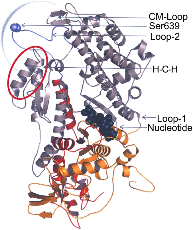
Fig. 6.
Homology model of the motor domain of Acanthamoeba myosin II. The motor domain shown in ribbon representation is based on the X-ray structure of Dictyostelium myosin II (Protein Data Bank ID, 2XEL) encompassing Met1 to Gln788.The N-terminal 25-kDa domain (red), central 50-kDa domain (gray), and C-terminal 20-kDa domain (yellow) are highlighted. Loop 1 connects the 25-kDa and 50-kDa domains, and loop 2 (purple) connects the 50-kDa and 20-kDa domains. The positions of Ser639 (purple) in loop 2 and the nucleotide-binding site, the helix–loop–helix motif (H-C-H, red circle), and the cleft in the 50-kDa domain that closes when myosin binds to F-actin (blue semicircle) are indicated. The model was generated using Modeller (27) and visualized using PyMol Version 1.4.1, Schrodinger, LLC).

