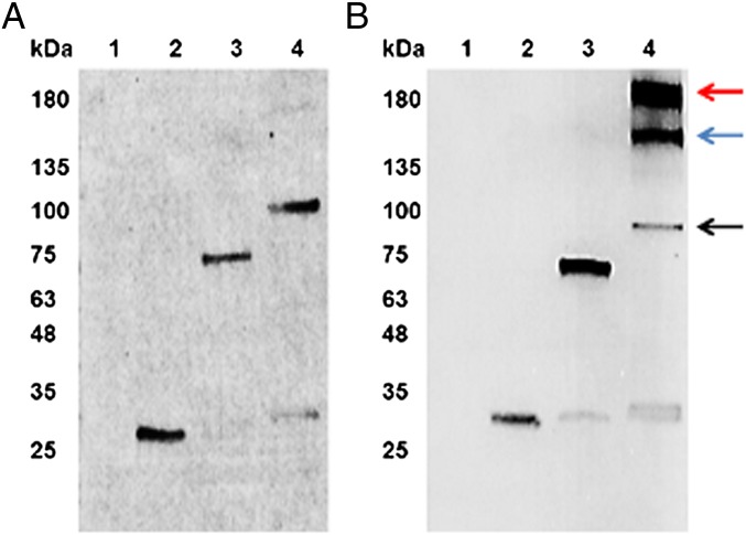
Fig. 3.
Western blots demonstrating the accumulation of immunotoxin proteins. (A) Samples (each 20 µg o.d) were separated on a SDS/PAGE gel under reducing conditions, transferred to a nitrocellulose membrane, and probed with an anti-Flag antibody that was conjugated with alkaline phosphatase. The lanes contain the following samples: lane 1, WT total protein; lane 2, αCD22; lane 3, αCD22PE40; lane 4, αCD22CH23PE40. (B) The identical samples were separated on a SDS/PAGE gel under nonreducing conditions to keep disulfide bonds intact. Once separated and transferred to a nitrocellulose membrane, samples were probed with an anti-Flag antibody conjugated with alkaline phosphatase and visualized on the nitrocellulose membrane. The black arrow indicates monomeric αCD22CH23PE40; the red arrow indicates αCD22CH23PE40 that has formed a homodimer which is indicative of an assembled antibody; the blue arrow indicates the formation of an assembled product between αCD22CH23PE40 and a degradation product lacking an scFv binding domain. This result demonstrates that algae produce αCD22CH23PE40 as a dimer, making it a divalent protein containing two exotoxin A molecules.

