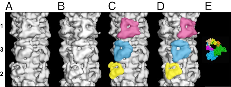
Fig. 1.
(A–D) Surface views of the final 3D reconstruction of the human cardiac myosin filament obtained by single-particle EM analysis and displayed using PyMOL showing a length of a full 429-Å repeat. The reconstruction is shown in two views (A and C; B and D) related by a 25° rotation about the filament axis, such that in A and C, a myosin head pair on level 1 is facing the viewer, and in B and D, a myosin head pair on level 3 is facing the viewer. Note that the two views in A and C and in B and D are quite distinct because of the different perturbations in the crowns. The triangular-shaped configurations for the three head pairs on each crown level are shown in yellow, blue, and pink for levels 2, 3, and 1, respectively, in C and D. The bare zone is at the bottom of the map in all of the four views. (E) Crystal structure for the head pair (11). The blocked and free heads are color-coded as in refs. 9–11. Blocked head: motor domain, green; essential light chain, orange; regulatory light chain, yellow. Free head: motor domain, cyan; essential light chain, pink; regulatory light chain, beige.

