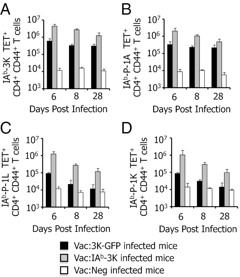
Fig. 2.
CD4 T-cell populations reactive to P-1L and P-1K in 506β mice are poorly expanded and not maintained in the spleens of mice infected with Vac:3K–GFP. Mice (506β) were infected with Vac:3K–GFP (black bars), Vac:IAb–3K (gray bars), or Vac:Neg (white bars). On days 6, 8, and 28 postinfection, 3K-reactive (A), P-1A–reactive (B), P-1L–reactive (C), and P-1K–reactive (D) CD4+ CD44+ T cells were quantified by staining with 1 μg/mL MHC tetramer. Data are the average of four (day 6), six (day 8), or five (day 28) total mice per group.

