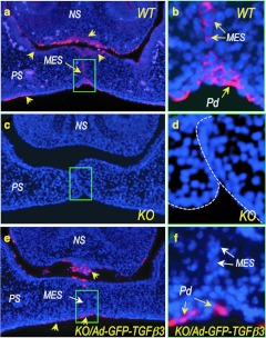
Figure 5.
Immunofluorescence localization of endogenous and virally expressed TGFβ3 in E14.5 mouse palatal shelves. (a) Endogenous TGFβ3 is detected in the palatal shelf (PS) epithelium on both oral and nasal sides, in the base of the nasal septum (NS), and the medial epithelial seam (MES) (yellow arrows) in wild-type (WT) mouse. (b) A higher magnification of the MES region indicated with a green box in a shows TGFβ3 expression in all MES cells. (c,d) Uninfected Tgfβ3−/− mouse palate shows complete absence of endogenous TGFβ3. (e) A rescued Tgfβ3−/− mouse palate with Ad-GFP-TGFβ3 (injected at E12.5 and harvested at E14.5) shows virally expressed TGFβ3 in the superficial layer of the palatal shelf and nasal septum epithelium (yellow arrows), but not in the MES (a white arrow). (f) A higher magnification of the green-boxed area in e confirms that the Ad-GFP-TGFβ3 transduced peridermal cells (Pd) are absent in the MES (white arrow). KO, knockout.

