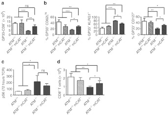
Figure 4.
Analysis of CD8+ T cell memory-differentiation phenotypes in vivo and in vitro. (a) Analyses of LCMV-specific Gp33+ splenic CD8+ T cells on day 30 post-LCMV infection of ATM+/+ (white-filled bar, n = 10), ATM+/+ mCAT+ (white filled with black stripes, n = 7), ATM−/− (gray-filled bar, n = 10), and ATM−/− mCAT+ (gray filled with black stripes, n = 9) mice. (b) Percent CD62Lhi, KLRG1+ (effector-memory TEM) and CD127+ (central memory, TCM) GP33+ CD8+ T cells from the mice analyzed in a. (c) Phospho-S6 MFI of splenic CD8+ T cells 72 hours post-TCR activation in vitro and (d) number of TCR-activated CD8+ T cells after additional 72 hours of IL-15 treatment from ATM+/+ (white-filled bar, n = 5), ATM+/+ mCAT+ (white filled with black stripes, n = 4), ATM−/− (gray-filled bar, n = 5), and ATM−/− mCAT+ (gray filled with black stripes, n = 5) mice. Statistical significance is denoted by *(<0.05), **(<0.005), ***(<0.0005) or ns (not significant). IL, interleukin; LCMV, lymphochoriomeningitis virus; MFI, median fluorescence intensity; TCR, T cell receptor.

