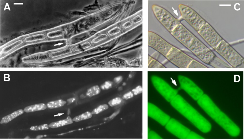
Figure 4 .
Conidia of Pyrenophora tritici-repentis connected by anastomosis bridges. (A) Phase-contrast microscopy of conidial anastomosis in Ptr. (B) The same conidia shown in A stained with DAPI and visualized under an ultraviolet filter to image nuclei; Note the loss of nuclei from the upper cell. (C) Nomarski optics (D) and fluorescence microscopy of anastomosed conidia of Ptr expressing green fluorescent protein. Arrows indicate the anastomosis bridges. Scale bars = approximately 5 µm.

