
Figure 3. Self-renewal and multipotency of Sox2+ stomach stem cells.
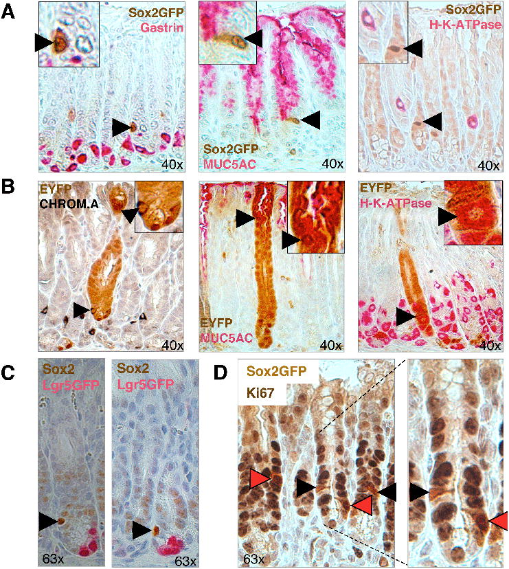
(A) Co-staining for Sox2-GFP and gastric markers on pylorus sections. Sox2-GFP+ cells (arrowheads) do not express differentiated cell markers gastrin (enteroendocrine cells), MUC5AC (mucus cells) or H-K-ATPase (parietal cells). (B) Co-staining of lineage tracing sections of Sox2-CreER; ROSA26-lsl-EYFP mice with differentiation markers in glandular stomach. Co-staining (arrowhead) was seen between EYFP and Chromogranin A (enteroendocrine cells) and MUC5AC (mucus cells) in the pylorus and with H-K-ATPase (parietal cells) in the corpus-pylorus transition zone. (C) Co-localization analysis for Sox2 and Lgr5-EGFP in pylorus. Sox2 (arrowhead) and Lgr5-EGFP (pink) IHC signals appear to mark different cells in pyloric glands. (D) Co-localization analysis between proliferation marker Ki67 and Sox2-GFP. Black arrowheads depict Ki67+ cells, red arrowheads depict Ki67+Sox2-GFP+ cells. Original magnifications as indicated. See also Suppl. Figure 4.