
Figure 4. Sox2-GFP+ testis cells reconstitute spermatogenesis in infertile mice.
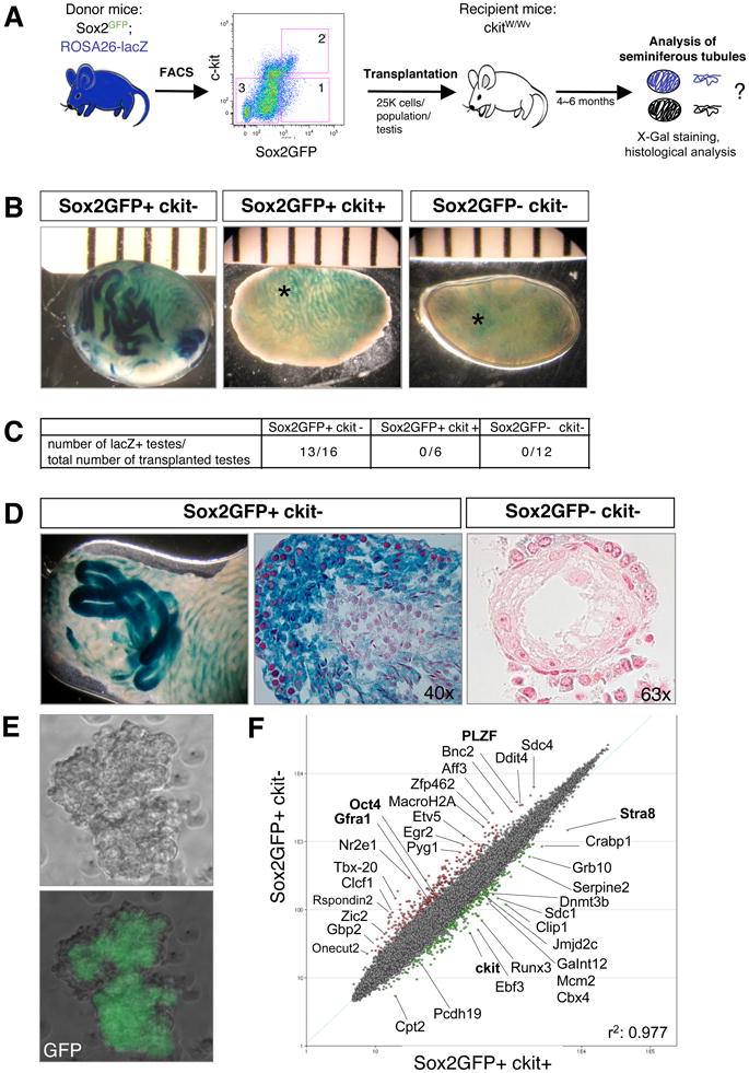
(A) Experimental outline. Sox2GFP animals were crossed with ROSA26-lacZ animals to enable tracking of transplanted donor cells. Sox2GFP+ckit− cells (1), Sox2GFP+ckit+ cells (2), and Sox2GFP−ckit− cells (3) from the testes of 2-week-old animals were transplanted into the vas deferens of germ cell-depleted, infertile ckitW/Wv recipient mice and testes analyzed for beta-gal activity after 6 months. (B) X-gal stain of transplanted ckitW/Wv testis 6 months after transplantation with the indicated cell populations. Light blue signal (asterisk) reflects background staining of the interstitium. (C) Table summarizing repopulation experiments. (D) Whole mount X-gal staining (left image) and paraffin sections of repopulated seminiferous tubules counterstained with neutral red (center image). Note the presence of immature spermatogonia at periphery and mature sperm in center of tubules transplanted with Sox2GFP+ckit− cells and lack of spermatogenesis upon transplantation of Sox2GFP−ckit− cells (right image). (E) Scatter plot of gene expression profiles comparing Sox2GFP+ckit− and Sox2GFP+ckit+ testis cells. Green dots depict genes with 2-fold and higher expression in Sox2GFP+ckit+ cells, red dots depict genes 2-fold and higher expression in Sox2GFP+ckit− cells. (pairwise analysis, two-fold change, t-test P=0.05, Benjamini and Hochberg correction.) Previously characterized spermatogonial stem cell genes are shown in bold. (F) Phase contrast and GFP images of a spermatogonial stem cell line derived from Sox2-GFP mice (passage 10). See also Suppl. Figure 5.