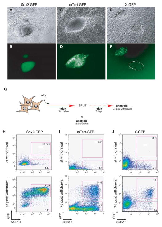
Figure 3. Timing of Sox2-GFP, mTert-GFP, and X-GFP reactivation during reprogramming.
(A–F) Brightfield and GFP images of iPS cells derived from Sox2-GFP (A,B), mTert-GFP (C,D), and X-GFP (E,F) fibroblasts. The dotted circle in (F) indicates a colony entirely GFP−, possibly due the loss of one X chromosome. (G) Experimental outline to determine temporal relationship of SSEA-1 expression, GFP marker activation and the appearance of doxycycline-independent iPS cells. Fibroblasts from the different GFP reporter lines were infected with the inducible lentiviruses, cultured in the presence of doxycycline for 10–12 days, and then split equally into two fractions: one fraction was immediately analyzed by flow cytometry and another fraction was analyzed after seven days of culture without doxycycline. (H–J) FACS plots showing GFP and SSEA-1 expression in the cultures at the time of doxycycline withdrawal (upper panels) and at 7 days post-withdrawal (lower panels). Sox2-GFP cells were analyzed with a FACSAria while mTert-GFP and X-GFP cells were analyzed with a FACSCalibur.

