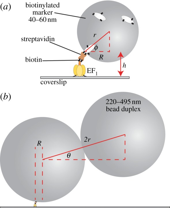
Figure 1.

The experimental set-up for observing the rotation of markers attached to surface-immobilized EF1 molecules. (a) A single bead (d = 2r = 40 nm shown) can only reach a minimum gyration angle ϕmin of sin(ϕmin) = 1 − h/r, where h is the height of the F1–streptavidin attachment point. (b) A bead duplex formed from two biotinylated polystyrene beads (d = 220 nm shown) will, in general, form a rise-angle of θ with the surface plane. The beads are attached via streptavidin (brown) and biotin (black lines). Cream spheroids represent biotinylated bovine serum albumin.
