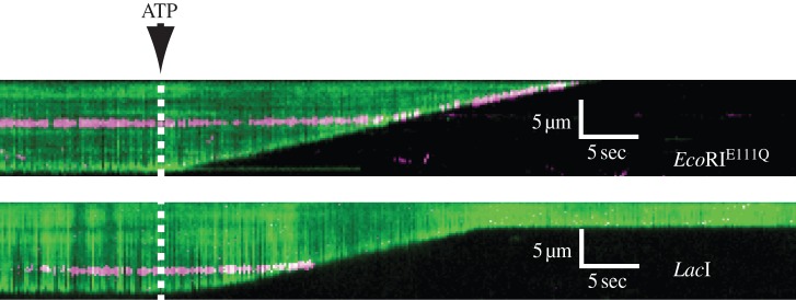
Figure 5.
The ATP-dependent directional translocase/exonuclease activity of RecBCD can be followed by tracking the shortening of a DNA strand as it is digested. The upper kymogram shows RecBCD encountering a magenta QD-labelled EcoR1 that is mutant for restriction activity. EcoR1 is pushed by RecBCD all the way to the nanofabricated barrier at the leading end of the DNA. The lower kymogram shows a similar experiment with the Lac1 repressor instead of EcoR1. Lac1 repressor is evicted from the DNA, and RecBCD continues translocating. Adapted with permission from Finkelstein et al. [52].

