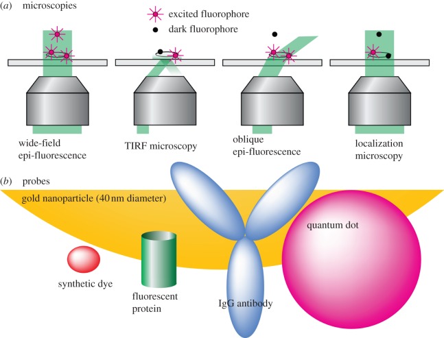
Figure 1.
Schematic of microscopies and probes used in single-molecule experiments. (a) Microscopies: different types of microscopy have different depths of excitation and different methodologies for reducing excess fluorescence. Shown are standard epi-fluorescence, TIRF, oblique angle epi-fluorescence and localization microscopies. (b) Probes: shown to approximate scale are the sizes of common fluorescent probes, including synthetic dyes (such as cyanine, Alexa or Atto dyes), fluorescent proteins, quantum dots (approx. 5–10 nm diameter) and gold nanoparticles (greater than 40 nm diameter). For comparison, an IgG antibody is also shown to approximate scale.

