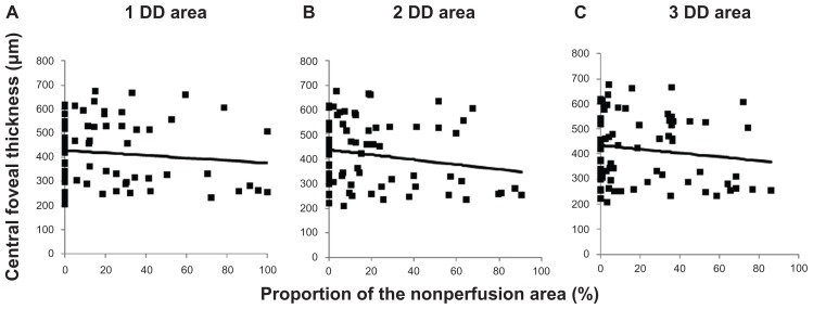
Figure 4.
Central foveal thickness is not associated with the percentage of the nonperfusion area. The plots show central foveal thickness and the percentages of the nonperfusion area in (A) the 1-disc diameter (r = 0.11, P = 0.361), (B) the 2-disc diameter (r = 0.18, P = 0.120), and (C) the 3-disc diameter (r = 0.14, P = 0.23) areas.

