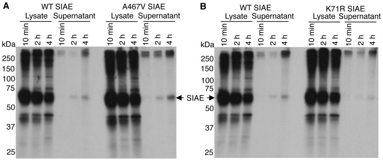
Figure 2. Metabolic labeling and pulse-chase analysis comparing the secretion of wild type SIAE with K71R and A467V SIAE proteins.
HEK 293T cells transfected with cDNAs encoding WT, K71R and A467V SIAE were pulse labeled with 35S-methionine and lysates and supernatants were immunoprecipitated with anti-Flag antibodies after 10 min, 2 hrs and 4 hrs of chase. Proteins were separated by SDS/PAGE and revealed by autofluorography. The position of molecular weight markers is indicated on the left in kilodaltons (kDa). (A) A comparison of wild type and A467V SIAE proteins. (B) A comparison of wild type and K71R SIAE proteins.

