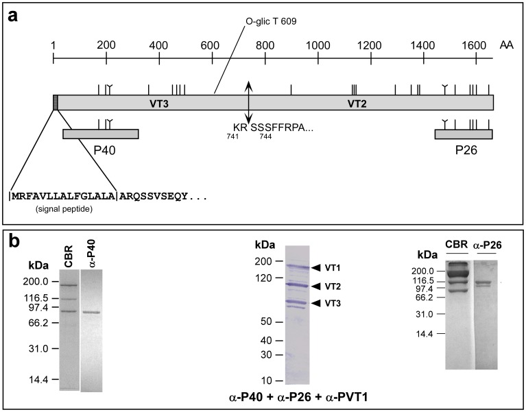
Figure 4. Positioning yolk polypeptides VT2 and VT3 in the vitellogenin precursor OTI-VIT-6.
(a) Graphical representation of OTI-VIT-6. The vertical lines show the position of each cysteine in the molecules; the “Y”-shaped lines show the position of the “CXXC” motif. The positions of the recombinant polypeptides expressed in E. coli are shown underneath the protein representation. The cleavage sites of the signal peptide and processing of the precursor were obtained using N-terminal sequencing of the mature VT2 and VT3. (b) Coomassie Blue-stained SDS-PAGE of purified O. tipulae vitellins (CBR) and the immunoblots using mouse anti-P40 (α-P40) and anti-P26 (α-P26) sera. The results from the analysis with all three anti-sera (α-P40+ α-P26+ α-PVT1) in the same blot are also shown. Each of the Western blotting strips represent a different SDS-PAGE experiment.

