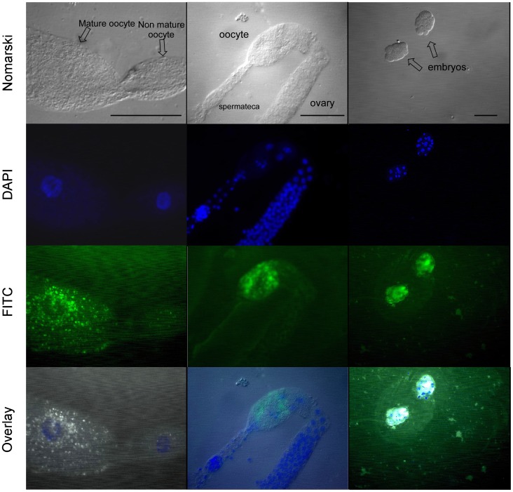
Figure 5. Immunofluorescence detection of yolk granules in the O. tipulae ovary and embryos using anti-P26 sera.
Worms were cut open with iridectomy blade over a gelatin-subbed slide, fixed and permeabilized. The slides were incubated with anti-P26 serum diluted to 1∶100 and subsequently incubated with FITC anti-mouse IgG in the same dilution. The slides were visualized using an Axiophot Zeiss photomicroscope, and photos were captured with epifluorescence (DAPI and FITC filters) or DIC for each field. The black scale lines in the first row (DIC/Nomarski photos) correspond to 50 µm.

