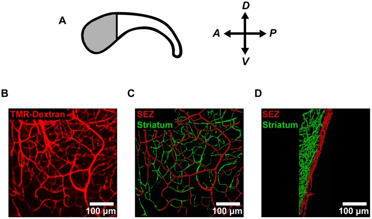
Figure 1. Confocal imaging reveals microvascular architecture in SEZ flatmounts.
(A) For analysis of vessel structure in the neural stem cell niche, we focused on the anterior region of the SEZ (shaded region) where the density of neural stem cells is highest. (B) The microvessels in the anterior SEZ were labeled with an intracardial injection of fluorescent tetramethylrhodamine-labeled dextran (TMR-Dextran) and imaged en-face. (C–D) Vessel segments were traced using image processing software and color-coded according to distance from the ependymal wall (SEZ = 0−20 µm; striatum >20 µm). Vessels are shown both en-face (C) and from the side (D).

