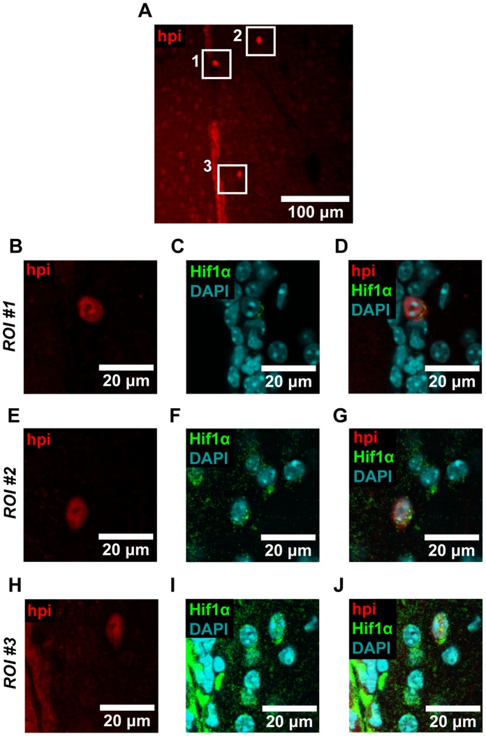
Figure 5. Hypoxyprobe-1 staining colocalizes with Hif1α.
(A) Coronal brain sections were imaged, and regions of interest (ROIs) where Hypoxyprobe-1 (hpi) staining was observed were selected for closer inspection. (B–J) We observed colocalization between Hypoxyprobe-1 and Hif1α in each of the three ROIs shown, and in cells located in the SEZ, striatum, and ependymal layer. Overall, we observed colocalization with Hif1α in 83% of the cells that exhibited bright Hypoxyprobe-1 staining (ncells = 18). Image data were processed with a Gaussian filter. Representative images are shown.

