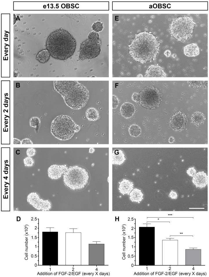
Figure 1. Growth of embryonic and adult OBSC neurospheres supplemented at different intervals with FGF-2 and EGF.
Embryonic OBSCs (A–D) and adult OBSCs (prepared from 4-, 6-, 7-, 12- and 15-month old mice; E–H) were grown as neurospheres, and FGF-2 and EGF (FGF-2/EGF) were added daily, every 2 days or every 4 days. On the day of passage (day 4 for embryonic and day 7 for adult OBSCs), the neurospheres were mechanically dissociated and the cell number was determined using the Trypan blue dye exclusion method. Images show representative E13.5 (A–C) and adult (E–G) neurospheres. The bar graphs show the average number of eOBSCs (D) and aOBSCs (H) in each condition. The results represent the mean ± SEM from 24-35 passages from 6 different cell cultures per condition. Decreasing the frequency of FGF-2/EGF addition reduced cell number and neurosphere size. *P<0.05, **P<0.01 and ***P<0.001 (Kruskal-Wallis test followed by post hoc analysis using Dunn´s multiple comparison test). The 36% reduction in eOBSC number in the C4 versus de Ctr condition (D) was statistically significant when the two average means were compared using the Student´s t test (P<0.01). Scale bars (G) = 121.02 µm.

