Figure 1.
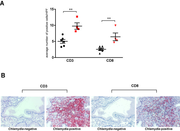
T cell infiltrates in endocervical tissue sections. (A) The number of CD3+, CD8+ cells in six C. trachomatis-negative (black) and four C. trachomatis-positive (red) endocervical tissues were assessed by immunohistochemistry. T cell counts represent the mean number of positive cells per high power field (HPF). Mean number of positive cells were derived from examination of twenty high power fields for each tissue sample. Statistical analysis was performed using Student’s t-test and confirmed by Wilcoxon Rank Sum test. Asterisks denotes p<0.01. (B) Representative C. trachomatis-negative and C. trachomatis-positive endocervical tissues stained with anti-CD3 and anti-CD8 antibodies. Red stain indicates CD3 or CD8 positive cells.

