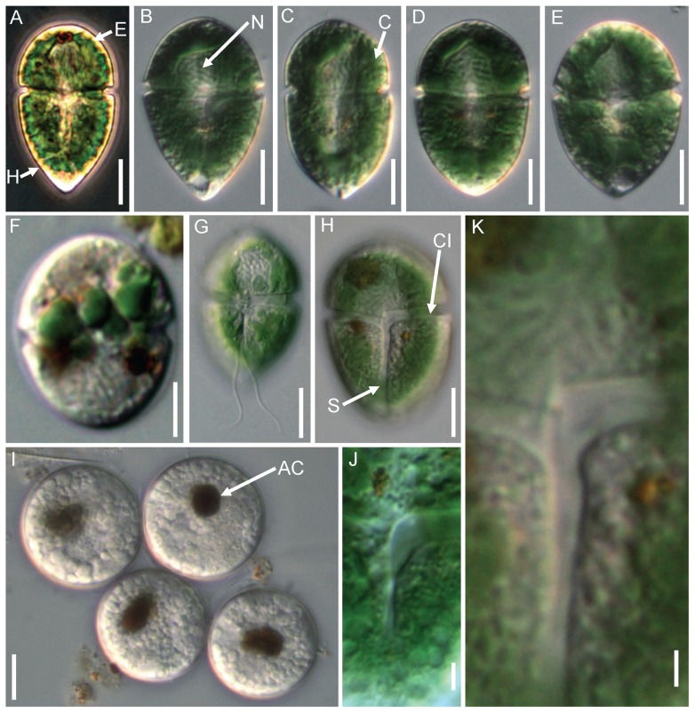
Figure 1. Micrographs of Gymnodinium eucyaneum.
Figs. A–E. Different cell shapes of the field samples. F Cells kept for 2∼4 weeks in the laboratory, showing that the chloroplast became smaller. G Ventral view showing the insertion of the flagella. H, J, K Ventral view showing the detail of the cingulum and sulcus. I Cysts each with a brownish accumulation of corpuscles. E: epicone; H: hypocone; N: nucleus; C: chloroplasts; CI: cingulum; S: sulcus; AC: accumulation of corpuscle. Scale bars: A–I = 10 μm; J–K = 2 μm.

