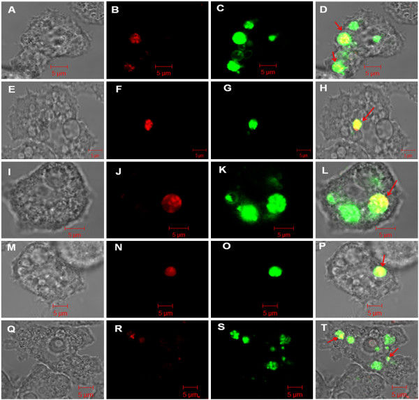
Figure 4.
Confocal microscopy analysis of stressed and non-stressed C. jejuni cells within acidic organelles of A. castellanii observed immediately after gentamicin treatment. Control C. jejuni (A-D), C. jejuni pre-exposed to osmotic stress (E-H), heat stress (I-L), hydrogen peroxide (M-P), or starvation stress (Q-T). The multiplicity of infection was 100:1 (bacteria:amoeba). (A, E, I, M, Q) differential interference contrast image; (B, F, J, N, R) C. jejuni stained with CellTracker Red; (C, G, K, O, S) acidic amoeba organelles stained with LysoSensor Green; (D, H, L, P, T) corresponding overlay. Scale bar = 5 μm.

