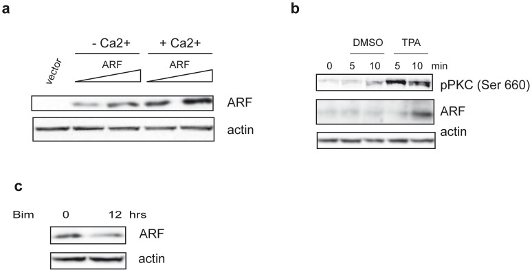
Figure 1. SDS-PAGE analysis of ARF protein levels.
a) HaCaT cells were transfected with an empty plasmid (vector) and increasing amount of p14ARF expression plasmid. Twenty-four hours after transfection cells were treated or not with 2mM Ca2+ as indicated. Cellular extracts were immunoblotted and analysed with anti His antibody to detect exogenous ARF levels and anti actin as loading control. b) HaCaT cells were treated with 10µM TPA for different time points. Protein extracts were subjected to WB with anti ARF (C-18) and an anti pPKC pan antibody that recognizes all the PKC isoforms phosphorylated at a carboxy-terminal residue homologous to serine 660 of PKC β II (activated pPKC). Actin was used as loading control. c) HaCaT cells were treated with 5µM Bisindolylmaleimide (Bim) for 12 hours (hrs) and analysed with anti ARF and anti actin antibodies.

