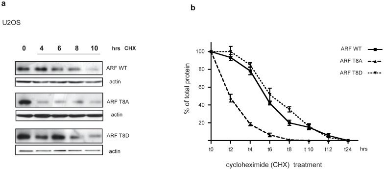
Figure 4. Protein turn-over analysis.
a) Half-life analysis of the wt and mutated p14ARF in human U2OS cells. Cells were transfected with p14ARF wt, T8A and T8D plasmids and treated with cycloheximide (CHX) for the indicated times two days after transfection. ARF and actin levels were analysed by WB of total extracts. b) The plot represents half-life analysis of wt and mutant ARFs. Band intensities were quantified by Image J analysis and actin normalized before being plotted in graph. The amount of protein at different time points is expressed as percentage of total protein, i.e. protein amount at t0. Each profile represents the mean of three independent transfections and WB experiments. Standard deviations are also shown.

