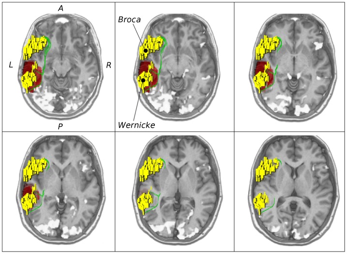
Figure 2. Validation on medical data.
Axial slices of MRI data in upward order with the Broca and Wernicke speech areas (yellow), fMRI activations (white and yellow) and ventral pathways along the inferior fronto-occipital fasciculus (IFOF) reconstructed by global search (GS) (green). Images show patient (female, 36 years old) with language lateralized to the left hemisphere and with a left temporal astrocytoma (WHO grade II, shown in red).

