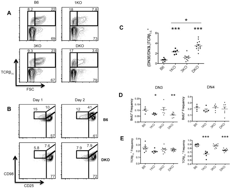
Figure 5. RasGRP1 KO and RasGRP1/3 DKO thymocytes display impaired proliferation of DN3 and inefficient transition from DN3E to DN3L. A.
Intracellular TCRβ (TCRβi.c.) by forward scatter (FSC) profiles of DN3 (CD4−CD8−Thy1.2+CD44−CD25+) from B6 (n = 8), 1KO (n = 6), 3KO (n = 9) and DKO (n = 12) thymi. B. Frequencies of DN3 (CD4−CD8−Thy1.2+CD44−CD25+) and DN4 (CD4−CD8−Thy1.2+CD44−CD25−) expressing intracellular TCRβ (TCRβi.c.). C. Ratio of frequencies of TCRβi.c. + DN3E/DN3L ((DN3E/DN3L)TCRβi.c. +). D. CD98 by CD25 profiles of Thy1.2+CD44− cells from 1 and 2 day DN3E-OP9-DL1 co-cultures; data are representative of 3 independent experiments. E. Frequencies of BrdU+ DN3 (CD4−CD8−Thy1.2+CD44−CD25+) and DN4 (CD4−CD8−Thy1.2+CD44–CD25−) from B6 (n = 5), 1KO (n = 5), 3KO (n = 6) and DKO (n = 7) mice injected with BrdU i.p. 2h prior to euthanasia. *p<0.05, **p<0.01 and ***p<0.001.

