Figure 1.
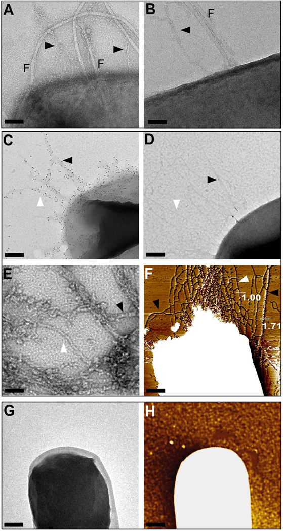
Assembly of pilus bundles on the surface of Bacilli. (A + B) Transmission electron micrographs of negatively stained wild-type B. cereus ATCC 14579. In both panels, B. cereus cells display surface exposed flagella (F) and pili (arrows) that associate as bundles. Bar = 50 nm. (C) Immuno transmission electron micrograph (TEM) of B. anthracis Sterne (pJB12). Pili were labeled using rabbit anti-BcpA antibodies followed by goat anti-rabbit IgG-10 nm gold conjugate to reveal fibers with variable diameter (bar = 100 nm). (D) B. anthracis Sterne (pJB12) was incubated with goat anti-rabbit IgG-10 nm gold conjugate alone. (E) Negative staining TEM micrograph of pili released B. anthracis Sterne srtA (pJB12) cultures. Single pili (white arrowhead), two-filament bundles (black arrowhead) pili, and higher order bundle assemblies were observed (bar = 20 nm). (F) Flattened atomic force microscopy (AFM) image of B. anthracis Sterne (pJB12) cells that elaborate pili with varying diameters as indicated by height measurements in nanometer (Bar = 100 nm). (G + H) Negative control, B. anthracis Sterne (pLM5) cells analyzed by negative staining TEM and AFM, respectively, did not display pili or pilus bundles at the surface of the cell (Bar = 100 nm).

