Figure 4.
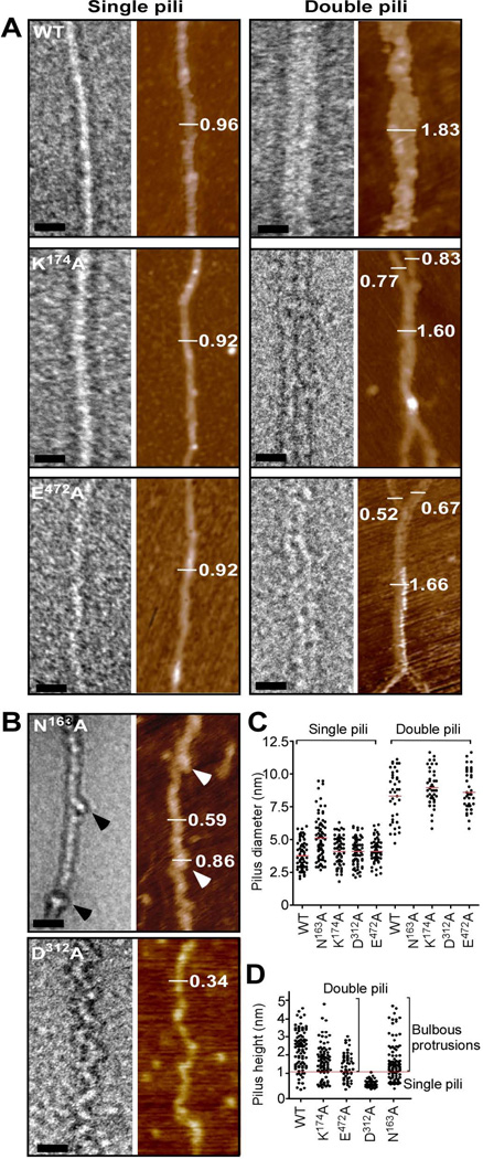
Electron microscopy and atomic force microscopy of wild-type and mutant pili. (A) A combination of TEM and AFM analysis of wild-type BcpA (WT), K174A, and E472A pili. TEM images were obtained of single and double filament pili negatively stained with 2% uranyl acetate (bar = 20 nm) and by AFM (mocha color). Single pili assembled from wild-type (WT), K174A and E472A mutant BcpA assume a straight morphology, while double filaments represent a close association of two pili that may intertwine. The visualization of single and double filament pili by TEM was confirmed by AFM as indicated by height measurements in nanometer. (B) Pili assembled from N163A variants harbor bulbous protrusions (arrowheads) in regular intervals along an otherwise straight filament. Mutant D312A pili appear as filaments with zig-zag or undulating deformations. Both pilus variants were only observed as single filaments. (C) Quantification of pilus diameter from TEM micrographs of pilus extracts of at least 2 independent experiments. Each dot represents a measurement of the diameter of one pilus. Multiple measurements were taken along >20 pili. (D) Quantification of pilus height on flattened AFM images of pilus extracts of at least 2 independent experiments. Each dot represents a height measurement of one pilus and multiple measurements were taken along >20 pili.

