
Fig. 4. Neutralization mechanisms of CR8033, CR8071 and CR9114.
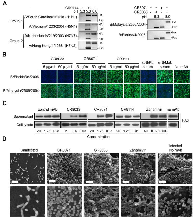
(A) CR9114 protects HAs of group 1 (A/South Carolina/1/1918 (H1N1) and A/Vietnam/1203/2004 (H5N1)) and group 2 (A/Netherlands/219/2003 (H7N7) and A/Hong Kong/1/1968 (H3N2)) influenza A viruses from the pH-induced protease sensitivity associated with membrane fusion. Exposure to low pH converts the HAs to the post-fusion state, rendering them sensitive to trypsin digestion (lane 1 vs. 3), but CR9114 prevents this conversion, retaining the HA in the protease-resistant, pre-fusion form (lane 2). CR8033 and CR8070 do not prevent this conversion at low pH (right panel) (N=4). (B) Expression of influenza NP in monolayers of MDCK cells 16-18 hrs after inoculation with B/Florida/4/2006 or B/Malaysia/2506/2004 viruses that were pre-incubated with CR8033, CR8071, CR9114, or polyclonal sheep sera directed against B/Florida/4/2006 or B/Malaysia/2506/2004, as indicated. NP expression is determined by immunofluorescence. Representative images of three independent experiments are shown. (C) Immunoblots of uncleaved HA (HA0) detected in the lysate and supernatant of MDCK cells infected with B/Florida/4/2006 virus and subsequently (from 3 to 20 hours post infection) incubated with different concentrations of antibodies or zanamivir as indicated. HA0 was detected using rabbit serum against B/Jiangsu/10/03 (Yamagata lineage). Concentrations are in μg/ml and μM for antibodies and zanamivir, respectively. Results from one representative of two independent experiments are shown. (D) SEM images of the surface of MDCK cells infected with B/Florida/4/2006 virus and subsequently (from 3 to 20 hours post infection) incubated with CR8071 (10 μg/ml), CR8033 (2.5 μg/ml), or zanamivir (60 μM). Representative images of three independent experiments are shown. Scale bar 1 μm.