
Fig. 3.
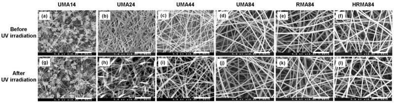
Scanning electron micrographs of electrospun unmodified methacrylated alginate (UMA), RGD-modified methacrylated alginate (RMA), as well as heparin and RGD-modified methacrylated alginate (HRMA) nanofibres (prior to PEO extraction) (a–f) before and (g–l) after cross-linking using UV irradiation. Images represent: (a and g) UMA14 (b and h) UMA24 (c and i) UMA44, (d and j) UMA84, (e and k) RMA84, and (f and l) HRA84. Scale bars represent 3 μm.