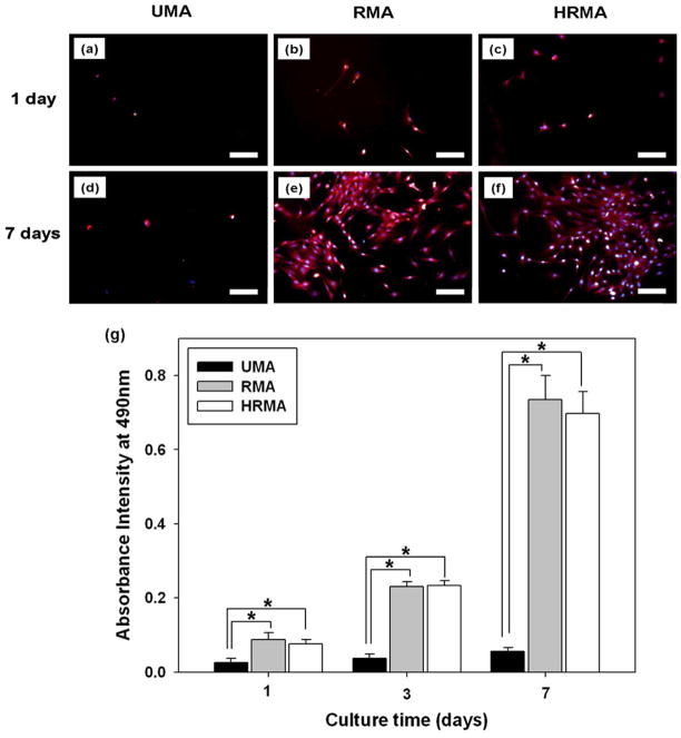
Fig. 8.
(a–f) Fluorescence photomicrographs of HDFs cultured on photo-cross-linked PEO-extracted UMA84, RMA84, and HRMA84 nanofibre scaffolds stained with rhodamine-phalloidin and DAPI. Scale bars represent 100 μm. (g) MTS assay of HDFs cultured on these nanofibre scaffolds for 1, 3 and 7 days. *p < 0.05.

