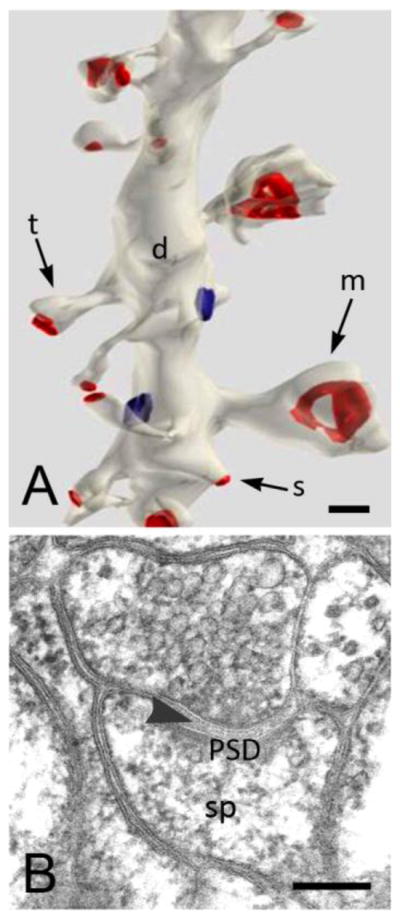
Figure 1. Appearance of dendritic spines in the electron microscope.

A. 3D reconstruction of serial thin sections (from stratum radiatum of rat CA1 shows stubby (s) spines on the same segment of a dendritic shaft as spines with thin (t) and mushroom (m) shapes. Synaptic contacts have been colorized in red. Image is from http://synapses.clm.utexas.edu/anatomy/compare/compare.stm, reprinted with kind permission from J. Spacek. B. Micrograph of a thin (70 nm) section shows a typical mushroom-shaped spine from the rat hippocampus. Note the presynaptic terminal with synaptic vesicles, the synaptic cleft (arrowhead) and the postsynaptic density (PSD). The filamentous material in the spine head (sp) represents the actin cytoskeleton. Scale bars: 200 nm
