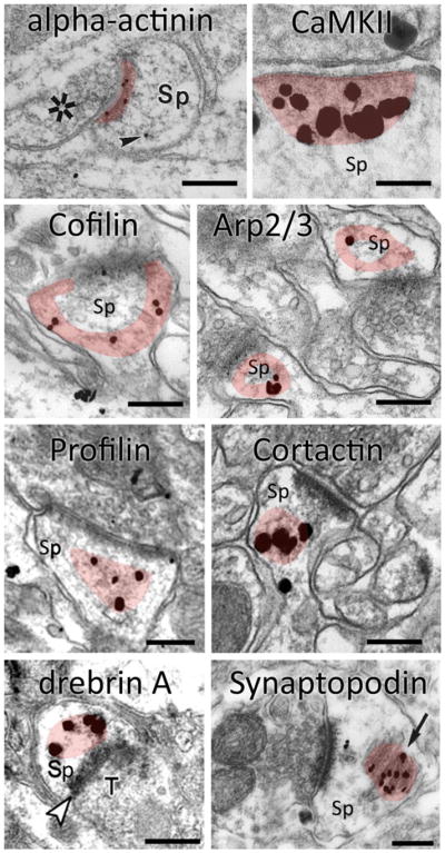
Figure 3. ABP content of spines, revealed by immunogold.

Hippocampal spines (Sp) labeled with immunogold for eight different actin-binding proteins. (Images adapted with permission from [109] (α-actinin), [116] CaMKII, [137] (cofilin), [149] (Arp2/3 complex),[163] (profilin), [157] (cortactin), [174] (drebrin) and [125] (synaptopodin). Shaded area represents the zone of concentration, for each protein. Scale bar: 200 nm.
