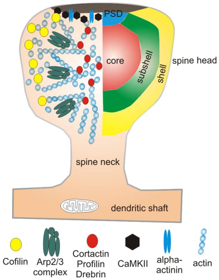
Figure 4. ABP microdomains in the spinoskeleton.

Proteomic studies of forebrain synapses have identified a sizable number of ABPs in the biochemically-defined PSD [110, 182, 183]. Some of these may be contaminants, but several, including α-actinin (blue double ovals) and CaMKII (black hexagons), have been demonstrated with immuno-EM to lie within the morphologically-defined PSD. Imaging studies reveal that actin (small blue circles) is more dynamic in the shell of the spine than in the center, suggesting a functional gradient of activity, from shell to core. Consistent with these data, cofilin (yellow circles) — a protein responsible for depolymerization of filaments — is heavily concentrated in this shell domain. The presence of a distinct “subshell” microdomain within the spinoplasm is suggested by the accumulation of the Arp2/3 complex (green composites), which mediates filament branching. The center (“core”) of the spine contains a relatively stable pool of actin. A heterogeneous pool of ABPs, including cortactin, profilin and drebrin (red circles), concentrate in this core microdomain.
