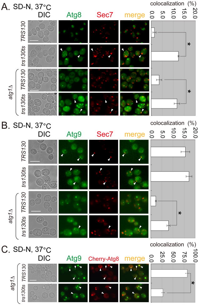
Figure 5. Atg8 and Atg9 are mislocalized in trs130ts mutant cells under starvation at the permissive temperature.
WT and trs130ts mutant cells (with ATG1 or atg1Δ) carrying the indicated fluorescent fusion proteins were grown in starvation conditions as in Figure 1A and examined for fluorescence. (A) Atg8 was partly colocalized with Sec7-DsRed (trans Golgi marker) in trs130ts mutant cells at 37°C after starvation. (B) Atg9 was partly colocalized with Sec7-DsRed in WT and trs130ts mutant cells at 37°C after starvation. (C) Drastically reduced colocalization between Atg8 and Atg9 was observed in the trs130ts mutant in atg1Δ background at 37°C after starvation. Atg9-3XGFP tagged WT and trs130ts mutant cells in atg1Δ background were transformed with Cherry-Atg8 plasmid (CUP1 promoter, LEU2, 2′). Cells were grown as in Figure 1A for starvation with 25 mM CuSO4 to induce Cherry-Atg8. The percentage of colocalization was calculated as green puncta to red puncta in (A) and (B), and red puncta to green puncta in (C) for two repeated experiments for a total of about 500 cells in ten fields. Results are presented as average percentage with standard deviation (right). Bar, 7 μm. Asterisks indicate P < 0.001 as highly significant.

